займы на карту
Search
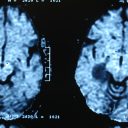
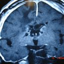
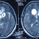
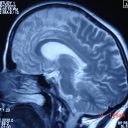
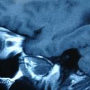
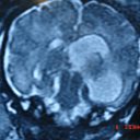
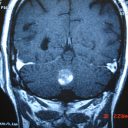
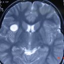
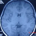
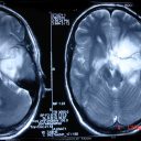
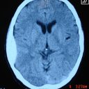
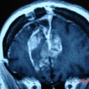
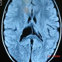
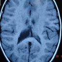
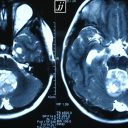
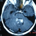
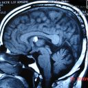
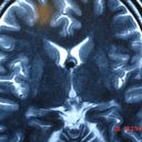
Brain – infiltrative tumor of deep parietal, DWI (2)
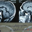
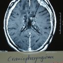
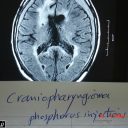
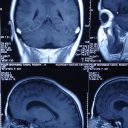
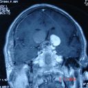
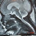
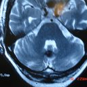
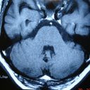
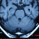
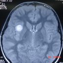
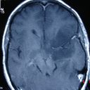
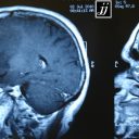
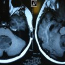
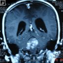
Brain – Craniopharyngioma phosphorous injection and
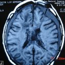
Brain – Craniopharyngioma phosphorous injection and
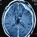
Brain – Craniopharyngioma phosphorous injection and
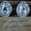
Brain – Craniopharyngioma phosphorous injection and
Brain – Craniopharyngioma phosphorous injection and
Brain – Craniopharyngioma phosphorous injection and
Brain – Craniopharyngioma phosphorous injection and
Brain – Craniopharyngioma phosphorous injection and
Brain – Craniopharyngioma phosphorous injection and
Brain – Craniopharyngioma phosphorous injection and
Brain – Craniopharyngioma phosphorous injection and
Brain – Craniopharyngioma phosphorous injection and
Brain – Craniopharyngioma phosphorous injection and
Brain – Craniopharyngioma phosphorous injection and
Brain – Craniopharyngioma phosphorous injection and
Brain – Craniopharyngioma phosphorous injection and
Brain – Craniopharyngioma phosphorous injection and
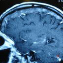
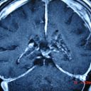
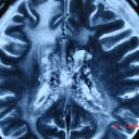
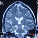
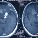
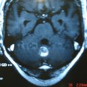
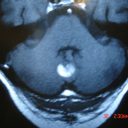
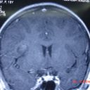
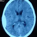
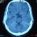
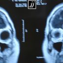
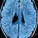
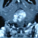
Brain – central skull base artifact from dental brid
Brain – central skull base artifact from dental brid
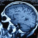
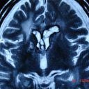
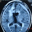
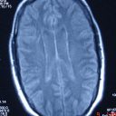
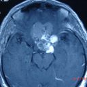
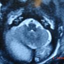
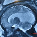
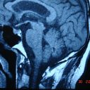
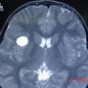
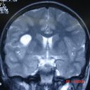
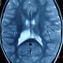
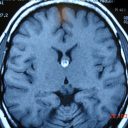
Brain- Brain – Craniopharyngioma ( T2)
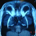
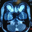
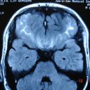
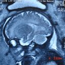
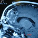
Brain- Brain- Brain – Frontal bossing and dolichocep
Brain- Brain- Brain – Frontal bossing and dolichocep
Brain- Brain- Brain – Frontal bossing (2)
Brain- Brain- Brain – Frontal bossing
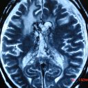
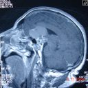
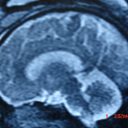
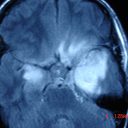
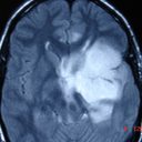
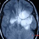
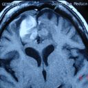
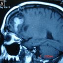
Brain- Brain- Brain – Craniopharyngioma (T2)
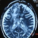
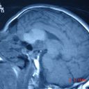
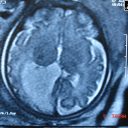
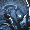
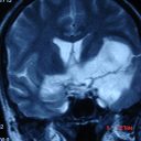
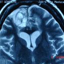
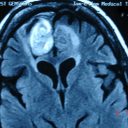
Brain- Brain- Brain – Craniopharyngioma (T1)
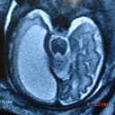
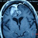
Brain- Brain- Brain – Craniopharyngioma (6)
Brain- Brain- Brain – Craniopharyngioma (5)
Brain- Brain- Brain – Craniopharyngioma (4)
Brain- Brain- Brain – Craniopharyngioma (3)
Brain- Brain- Brain – Craniopharyngioma
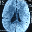
Brain – Bi frontal sub arachnoid hemorrhage by flair
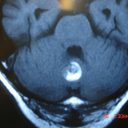
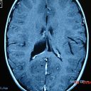
Brain – Anterior dental artifact coming from dental
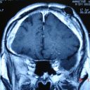
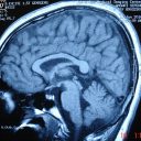
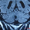
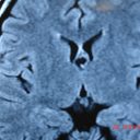
Brain – Anatomy
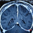
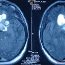
Fetus – bi lateral COM, brain Atrophy and hydrocepha
Fetus – bi lateral COM, brain Atrophy and hydrocepha
Fetus – bi lateral COM, brain Atrophy and hydrocepha
Fetus – bi lateral COM, brain Atrophy and hydrocepha
Fetus – bi lateral COM, brain Atrophy and hydrocepha
Fetus – bi lateral COM, brain Atrophy and hydrocepha
Fetus – bi lateral COM, brain Atrophy and hydrocepha
Fetus – bi lateral COM, brain Atrophy and hydrocepha
Fetus – bi lateral COM, brain Atrophy and hydrocepha
Fetus – bi lateral COM, brain Atrophy and hydrocepha
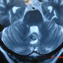
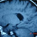
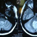
Brain – Vermis Cavernoma, T2 axial
Brain – Vermis Cavernoma, T2 axial, with hemorrhage
Brain – Vermis Cavernoma, T2 axial (3)
Brain – Vermis Cavernoma, T2 axial (2)
Brain – Vermis Cavernoma, T1
Brain – Vermis Cavernoma, T1 sagittal
Brain – Vermis Cavernoma, T1 sagittal after hemorrha
Brain – Vermis Cavernoma, T1 axial, with hemorrhage
Brain – Vermis Cavernoma, T1 axial, with hemorrhage
Brain – Vermis Cavernoma, T1 axial, with hemorrhage
Brain – Vermis Cavernoma, T1 axial with hemorrhage
Brain – Vermis Cavernoma, coronal FLAIR
Brain – Vermis Cavernoma, coronal FLAIR (2)
Brain – small hematoma, MRI, T2
Brain – small hematoma, MRI, T2 (3)
Brain – small hematoma, MRI, T2 (2)
Brain – small hematoma, MRI, T1
Brain – small hematoma, MRI, T1 (4)
Brain – small hematoma, MRI, T1 (3)
Brain – small hematoma, MRI, T1 (2)
Brain – small hematoma, MRI, sagittal T2
Brain – small hematoma, MRI, coronal T2
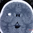
Brain – small hematoma, CT
Brain – Slow Growing infiltrated non enhancing tumor
Brain – Slow Growing infiltrated non enhancing tumor
Brain – Slow Growing infiltrated non enhancing tumor
Brain – Slow Growing infiltrated non enhancing tumor
Brain – Slow Growing infiltrated non enhancing tumor
Brain – Slow Growing infiltrated non enhancing tumor
Brain – Right parietal epidural hematoma
Brain – Right parietal epidural hematoma (5)
Brain – Right parietal epidural hematoma (4)
Brain – Right parietal epidural hematoma (3)
Brain – Right parietal epidural hematoma (2)
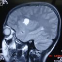
Brain – right frontal tumor, crossing falx
Brain – right frontal tumor, crossing falx, T2
Brain – right frontal tumor, crossing falx, GD T1
Brain – right frontal tumor, crossing falx, GD T1 (5
Brain – right frontal tumor, crossing falx, GD T1 (4
Brain – right frontal tumor, crossing falx, GD T1 (3
Brain – right frontal tumor, crossing falx, GD T1 (2
Brain – right frontal meningiomas
Brain – right frontal meningiomas (3)
Brain – right frontal meningiomas (2)
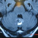
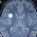
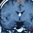
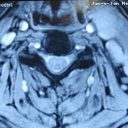
Brain – Right choroid plexus cyst
Brain – Right choroid plexus cyst (7)
Brain – Right choroid plexus cyst (6)
Brain – Right choroid plexus cyst (5)
Brain – Right choroid plexus cyst (4)
Brain – Right choroid plexus cyst (3)
Brain – Right choroid plexus cyst (2)
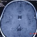
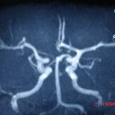
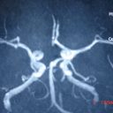
Brain – MRA, ACAs originate from left side wilis
Brain – MRA, ACAs originate from left side wilis (2)
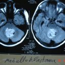
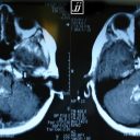
Brain – Medulloblastoma, T2
Brain – Medulloblastoma, T2, Hydrocephaly
Brain – Medulloblastoma, T2 (2)
Brain – Medulloblastoma, T1
Brain – Medulloblastoma, T1, Tonsillar herniation
Brain – Medulloblastoma, T1 GD
Brain – Medulloblastoma, T1 GD (5)
Brain – Medulloblastoma, T1 GD (4)
Brain – Medulloblastoma, T1 GD (3)
Brain – Medulloblastoma, T1 GD (2)
Brain – Medulloblastoma, T1 (4)
Brain – Medulloblastoma, T1 (3)
Brain – Medulloblastoma, T1 (2)
Brain – Medulloblastoma, Flair
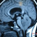
Brain – Lipoma of Mid line, sagittal T1
Brain – Lipoma of Mid line, FLAIR
Brain – lipoma of Mid line, Coronal T1
Brain – lipoma of Mid line, Coronal T1 (2)
Brain – lipoma of Mid line, Axial T2
Brain – Lipoma of Mid line, Axial T1
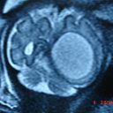
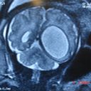
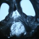
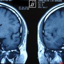
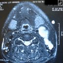
Brain – Left neck mass and Adenopathy
Brain – Left neck mass and Adenopathy, STIR T2
Page 192 of 235
« First
«
...
10
20
...
190
191
192
193
194
...
200
210
...
»
Last »